
The Breast Cancer Screenings
Like most forms of cancer, early detection plays an important role in the effective treatment of breast cancer—making routine breast cancer screenings a necessity. In recognition of Breast Cancer Awareness Month, the health experts at AVMC’s Antelope Valley Outpatient Imaging Center (AVOIC) are here to outline
Mammograms
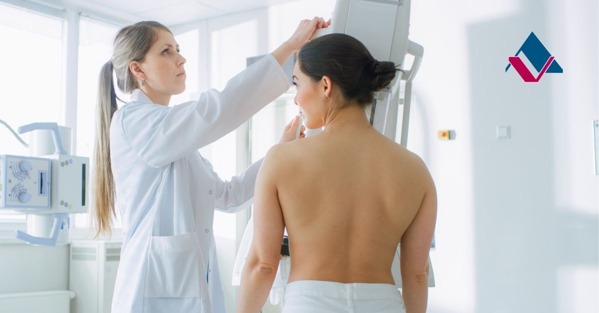
Mammograms are X-ray images of your breast tissue that are done as a form of breast cancer screening during well-woman visits with your primary care physician. When getting a mammogram done, you will stand in front of an X-ray machine where your breast will be placed on a plastic plate while another plastic plate will firmly press your breast tissue from above. After images have been taken from this view, it will be repeated to get a side view of your breast tissue.
Typically, women of average risk of developing breast cancer will begin getting mammograms around the age of 40. And should have them done annually.
Tomosynthesis (3D) Mammograms
Breast tomosynthesis, also known as a 3D mammogram, is an advanced form of mammography used to help doctors to detect breast cancer. This type of imaging uses low-dose x-rays to take a series of images of your breast tissue from different angles, which are then synthesized into a 3-dimensional image by the computer.
This effectively prevents any tissue overlap that may occur with traditional mammography, giving doctors a better view of your tissue and helping them to identify tumors.
Breast Ultrasound
Breast ultrasounds use sound waves to take images of your breast tissue, allowing your doctors to see certain changes in your breast tissue that may be difficult to see on a traditional mammogram. Typically, ultrasounds can be used to tell the difference between fluid-filled cysts and solid masses.
Breast ultrasounds can also be used during a biopsy procedure to ensure the accuracy of the needle being used to get a cell sample for testing
Self-Breast Exam
Between well-woman visits, it is important for women to become familiar with their own breast tissue and keep an eye out for any abnormalities. It is advised that women of all ages conduct self-breast exams at least once a month. When performing a self-exam, be sure to keep an eye out for the following::
- Lumps, bumps, or knots.
- Pain affecting the nipple or breast.
- Discharge other than breast milk.
- Flaking skin near the nipple.
- Dimpled skin.
- Changes in size or shape of the breast.
Breast Magnetic Resonance Imaging (MRI)
Breast magnetic resonance imaging (MRI) is typically done once breast cancer has already been detected in order to determine the size and location of cancer. In some instances, women with an increased risk of developing breast cancer will also have a breast MRI and an annual mammogram.
During a breast MRI, you lay face down on a table and your breasts will hang into an opening in the table so that they can be scanned without the need for compression. Your MRI technologist may also ask you to hold your breath in order to keep the imaging as clear as possible.
Specialty Care in Lancaster, CA
As a full-service radiology center, Antelope Valley Outpatient Imaging Center (AVOIC), is patient-focused and delivers high-quality imaging. Our knowledgeable staff is dedicated to providing our patients with heartfelt, compassionate care and medical excellence in four imaging centers around the Antelope Valley.
For questions or to make an appointment at the Antelope Valley Outpatient Imaging Center please call 661-726-6700. For more information about AVOIC please visit our website.

.2204261124304.jpg)